We come across a wide variety of cases in our routine OPD of a general hospital. Many a times a lot of cases need a logical thinking and application of certain techniques which probably are not described as gold standard protocols but we know that applying these can only lead us to the desired direction.
Few days back I came across one such case. It was easy to conclude but definitely needed experience and logical thinking and insight into anatomy.

INTRODUCTION OF THE CASE
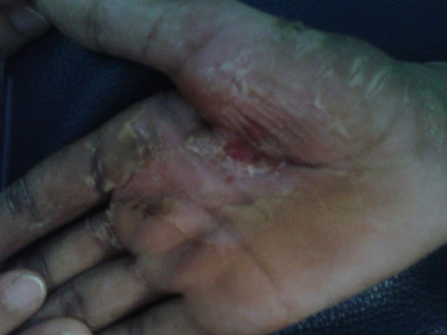
A male patient with glass cut injury over (rt) palm over first and second metacarpel region. Patient referred for EMG NCV studies by Plastic surgeon who wanted us to detect the exact site and extent of lesion.


Few days back I came across one such case. It was easy to conclude but definitely needed experience and logical thinking and insight into anatomy.

INTRODUCTION OF THE CASE
A male patient with glass cut injury over (rt) palm over first and second metacarpel region. Patient referred for EMG NCV studies by Plastic surgeon who wanted us to detect the exact site and extent of lesion.

CLINICAL EXAMINATION IN BRIEF
The scar on examination was adherent to the underlying structures especially entrapping the flexor tendon of index finger. Clinically minimal sensations present over index finger, minimal hypoaesthesia over middle finger. Normal sensations over thumb and ring fingers.
Patient was apprehensive to perform finger movements due to pain.
ELECTRODIAGNOSIS
Now we will try to logically appraise the rationale of each and every electrodiagnostic test performed with its interpretation
After the clinical assessment, NCV studies were begun.


- Motor Nerve Conduction Velocities both Median and Ulnar nerves were within normal limits with normal distal motor latencies and Compound motor action potential (CMAP) amplitudes.
- Latencies and amplitudes were bilaterally symmetrical and comparable.
- This suggest that the scar did not affect the motor component of (rt) Median nerve.


Above tables show many numbers with the titles which may sound different than the textbook ones and would for sure keep someone thinking as to what happened.
- Distal Sensory latencies and amplitudes both Ulnar nerves were bilaterally symmetrical and comparable which suggested that sensory component of Ulnar nerve was not involved.
Traditional Orthodromic Median Sensory Conduction involves stimulating from Index finger and recording from wrist.
- On the (rt) side, Sensory nerve action potential (SNAP) could not be elicited. This suggested that there was affection of Sensory branch of Median nerve on the right side.
Traditional viewpoint would confirm Median nerve injury and would interprete and report the same. But is this enough when we are talking about exact location and extent of injury?
To still go into the depth of the extent of injury, we need to re-evaluate the scar and once again understand the branching of the digital sensory branches of Median nerve
 |
| Add caption |

Looking to above two images the first one showing the branching pattern of the digital branch of Median nerve and the second one showing the site of scar. If we correlate the two, branch 1 going to the thumb would be spared. Branch 2 and 3 three going to index finger would be affected and that was the reason we could not elicit SNAP for Median nerve performed as a traditional technique. But the same Median nerve supplies Middle, ring and lateral half of ring finger as well as branch 4,5,6 in the figure showing nerve branching.
That is the need of today to think out of the box and perform SNCV of digital branches of Median nerve for thumb, middle and ring finger.
We could elicit SNAP for thumb, middle and ring finger for Median nerve. We performed Orthodromic technique with ring electrodes stimulating and surface electrodes placed on wrist recording.
- Distal sensory latencies of (rt) Median nerve for thumb, Middle and ring finger was nearly same.
- SNAP amplitude for middle finger was approximately 50% less than that of thumb and ring finger, though it was within normal limits.
Correlating this with the shape and location of the scar, probably branch number 2,3 and 4 would have been injured due to trauma.
This clinical and electrophysiological arguement was digestible to give a conclusion that there was affection of sensory branch only of the index finger and partial affection of the branch to middle finger.
Yet, as a protocol and to be confirmed that there was no motor involvement even to the minimal extent, EMG study was performed for two muscles, one muscle proximal to the scar Flexor Pollicis Brevis and one muscle distal to the scar 2nd Lumbrical

Though FPB has dual innervation, if Median nerve was denervated at the site of scar, it would definitely give signs of denervation like Fibrillation Potentials or Polyphasic Motorunit action Potentials.
Looking to the table, EMG study of both the muscles tested was normal once again proving with final confirmation that there was no motor involvement. Hence this case can be concluded as

The case looks very simple on first look. But frankly talking about interpreting it electrophysiologically, majority of electrophysiologist would stop at absence of SNAP with traditional technique and interpreting as Median nerve injury at the level of scar. The precision of interpretation of exactly saying it to be only for the branch of Median nerve for Index finger may not have any SO-CALLED RESEARCH EVIDENCE OR TEXT BOOK DOCUMENTATION. Nor does the technique employed to conclude the same has RESEARCH EVIDENCE. BUT THIS DEFINITELY HAS A CLINICAL CORRELATION WHICH IS PROBABLY MORE IMPORTANT THAN ANY TEXTBOOK, JOURNAL OR RESEARCH SAYING BECAUSE AFTER ALL MEDICAL SCIENCE IS FOR THE BETTERMENT OF THE PATIENT NOT FOR MERE DOCUMENTATION.
No comments:
Post a Comment