Sunday 20 December 2015

USING NCV STUDIES IN A ROUTINE PHYSIOTHERAPY CLINIC - A DIFFERENT WAY TO LOOK AT ELECTRODIAGNOSTIC STUDIES

Everyone of us aim to progress in life and dream to become a successful professional. We look high upon the professionals who have reached a certain height in their respective fields. But this doesnt come so easy. It is rightly said IF YOU DONT BUILD YOUR DREAMS, SOMEONE WILL HIRE YOU TO BUILD THEIRS. And to build our dreams apart from hard work we need a different thought process, innovations in the way we do our work and of course thorough knowledge of the subject with its application
A physiotherapist opening a new clinic will think of all equipments like electrotherapy equipments, exercise gadgets etc. But if said do buy one EMG NCV reply, he will give me this look


Somehow there is a phobia amongst physiotherapist regarding even looking at this equipment.
In our routine day to day clinical set up, we come across so many different varieties of patients ranging from DQ to radiculopathy and neuropathy. A physiotherapist specialized in Orthopedic Physiotherapy will have an orthopedic perspective in treating such patients. He will be restricted to muscles, joints, ligaments and manual therapy.The one specialized in Neurology will have his own perspective. He will restrict his thought process mainly to brain and spinal cord. And in this visualization, ultimately Peripheral Nervous System gets ignored and unexplored. That is the main reason why EMG NCV equipment and the concept itself has created a fear among physiotherapist. But let us see altogether different perspective to this which can create wonders to your patients

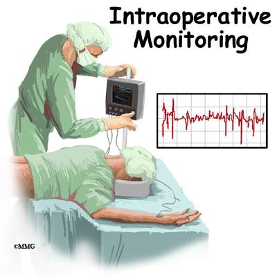
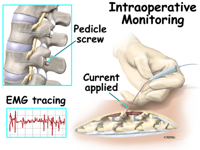
So many times we come across patients who inspite of being treated for days and months show little or no improvement. All our treatment modifications, techniques and advices prove worthless and we wonder now what.These are the times when EMG NCV equipment serves two purpose. One is it helps in further investigating the problem and secondly a therapist can charge more for performing a test. We may do mere NCV studies which does not require any certification. We may not give report as given in a electrophysiological studies. This can be mere one of our investigation just like Strength Duration Curve. But definitely helps.

Many neurologists adopt this. They screen the patient with NCV studies. Without releasing an official report but can come to a conclusion. We have seen so many Orthopedicians having X-ray machine in their clinic itself and so taking X-rays for the patient are just one step away. Why cant we have EMG NCV equipment?

Very few of us are aware about Pronator Teres syndrome, Ligament of Struther's syndrome which are entrapment of Median nerve at/around elbow which are often mistaken as Carpal tunnel Syndrome. Our neural tissue mobilization for such patients is focussed at wrist and hence inspite of putting all efforts, patient hardly improves. This is the time one may think of performing NCV study and reconfirm the diagnosis which can improve patient's outcome. Patients taking treatment for Tennis elbow or Lateral epicondylitis are one more example. All sort of electrotherapy, taping, manual therapy etc without much improvement leaves us in surprise and patient ultimately loses faith. Very few of us are aware of the fact that compression of Radial nerve between the heads of supinator traditionally called Supinator tunnel syndrome mimics Lateral epicondylitis. All symptoms usually are same as pain in wrist extension and weakness of wrist and finger extension which blindfold us to believe that this is due to pain which may not be the case. Simple NCV study can help detecting it and ultimately better patient outcome. Superficial Radial sensory nerve compression due to tight wrist bands can mimic radiculopathy. Patients take treatment of radiculopathy all in vain and ultimately they lose  faith in us. Sensory Radial conduction abnormality can help detect this. Suprascapular nerve neuropathy patient mistaken for Rotator cuff injury patient which again changes the treatment protocol and hence the patient outcome. Tarsal tunnel syndrome, radiculopathy and neuropathy have nearly same kind of sensory symptoms. Treating such wrongly diagnosed patients can definitely affect the clinical practice and patient - doctor trust and relationship. Simple NCV studies can help over come this serious problem. Such unknown entrapments are many. The list is beyond the description of this blog. But a newer perspective is definitely the aim of this blog. 

All it needs is a combined perspective of Orthopedics and Neurology with insight into Peripheral Nervous System. Thorough knowledge of anatomy, pathomechanics, pathology and of course good hands on skills and knowledge of NCV studies.

But this definitely does not need any certification as said by so many Medicos. All it needs is a newer way of seeing the patient. In India EMG NCV equipment are not so costly that one cannot afford to buy if he is making his own clinical set up. If it is yielding good patient outcome, that investment is worth doing.


Think Big, Work Hard, Think and implement something new that has a logical scientific base and one day you will have a machine that can produce what you have been dreaming of always.


Sunday 8 November 2015

EMG BIOFEEDBACK TRAINING - IN AN INDIAN CLINICAL SET UP - IS IT ACTUALLY THAT DIFFICULT?

There are two misconceptions that I see and I always want to clear them. 
One is students think that EMG biofeedback is too hard to understand. Ultimately they end up either leaving that topic in option or they just cram the topic to be able to write a short note in examination and forget the next day. 
The second misconception prevail among the qualified therapists. They always feel Clinical EMG equipment is too costly to purchase and they are not certified to perform the study and then it becomes a useless expense. Can anyone think that the same Clinical EMG equipment can be used for training weak muscles as EMG biofeedback? No doctor would try to create controversy saying we cannot perform this because muscle training is our forte and no one can deny that. We dont need those expensive gadgets to give biofeedback training. 

SURPRISED ?   

LETS SEE......

And before we see the clinical aspect let us understand the feedback loop in a very simplified manner.


The best example that one can give is of a small child when a parent is teaching him do some new task like pull-ups in the garden. And sitting in a corner we see the father's hands cheering him up and shouting, " YES, YOU CAN DO IT, SEE YOU HAVE DONE IT, ITS JUST THIS MUCH" and by these words gradually the child accomplishes this once difficult task. This is feedback. Furthermore once the child achieves a bit more, he himself sees the difference which his brain registers and then is further motivated to perform more and more until the task is accomplished.

Whenever we are giving strengthening exercises to the patients, we continuously give such encouraging commands to the patient to enable them perform better each day. This in itself is a feedback. When a patient performs a muscle contraction, electric energy is produced which is recorded by our electrodes and displayed on the screen. Hence patient can see and hear his own muscle performance. And since it involves a biological tissue, it is called biofeedback.

Using the same feedback for training a muscle using electromyography is Electromyography Biofeedback or EMG biofeedback. This doesnt mean this is too difficult a concept to understant and implement. All it needs is out of the box thinking and actually applying our learnt theory in the practical scenario.

Recent research evidences give controversial results, rather detrimental results regarding giving electrical stimulation for muscle strengthening. In such case, what is left for us to train such muscles?  Are only passive movements and active movements enough for rehabilitation? Can it give patient satisfaction? Rather than giving a controversial treatment just to satisfy the patient, why not think different and think new?

The same equipment that performs NCV and EMG studies can be used for this purpose. I know this leaves many of the readers in surprise. But simple. If we can see Motor Unit Action Potentials and Interferential Pattern of EMG studies in an equipment, why cant we use the same computer screen sequentially to see the progress of the patient, to let the patient see his progress and in a way get a feedback? Logical. Isn't it? If needle EMG study can be interpreted on the clinical EMG machine for diagnostic purpose, why can't we perform surface EMG for therapeutic purpose to train the muscle? This is EMG biofeedback. No one can create any controversy over this use of this equipment.

HOW TO SEE THE SCREEN


 The above figures take us back to our childhood days when we used to do maths. This reminds us of the graph paper wherein each square is one centimeter square area with each connecting horizontal and vertical line one centimeter. We used to adjust X-axis and Y-axis and plot a graph and actually enjoy doing it. We had to decide a unit like X-axis, 1cm= something and Y-axis 1cm=something and then plot a graph.


Now look at the above figure. Though it looks the same as the graph chart, is not our childhood graph. But it is the screen of the EMG equipment we use in clinical set up. They are so identical. Isn't it? Adjustments are also identical. The way we adjust X axis and Y axis in the graph, for the EMG screen, we adjust the horizontal screen and the vertical screen. Irrespective of using it for MUAP analysis or Biofeedback training, basic screen adjustments remain the same.The vertical line stands for the amplitude or the strength of contraction as measured in microvolts or millivolts depending upon patient's need. The horizontal line stands for time as measured in milliseconds. We can adjust the amplitude in the graph known as GAIN ( amplitude/cm) and time known as SWEEP ( sweep speed/sec)

In a graph, increasing or decreasing the units of X or Y axis changes the size of the graph. Similarly increasing or decreasing the gain and sensitivity will change the size of the graph seen on the EMG computer screen. This can be used as a feedback to train the patient.

While training a muscle, we adjust a gain. We tell the patient to perform the muscle action and see how much is the total amplitude of that action is. This gives us an idea of patient's capability of performance. We adjust the threshold accordingly. THRESHOLD is the maximum limit which we aim the patient should be able to do at that particular time. And we instruct the patient to perform the activity such that the MUAP crosses the threshold line


Above figure is an example of one such patient scenario graph. Red graph is the MUAP when a patient performs muscle contraction. "2"  which is seen on Vertical axis can be considered as Threshold and we instruct the patient to contract still strongly so that the red graph crosses the horizontal line passing through "2". Patient seeing the electric conversion of his own contraction as a graph performs still stronger contraction and ultimately puts in efforts to reach "2".
Progression can either be made by telling the patient to reach "3" or by increasing the gain. Once we increase the gain, graph size will be smaller and then once again we can instruct the patient to reach"2". On seeing the graph smaller, patient will definitely remember previous size and put in efforts to reach the previous day's target because patients know target and not the GAIN values. This is how progression made without any documented contraindications.

The same can be used to relax over active muscle aswell. Just reverse the sequence. Tell the patient to bring down the activity level, relax so that the graph falls below the threshold and progressed by reducing the gain. 

ELECTRODES USED - 

Surface electrodes are used. Two surface electrodes placed over the muscle to be trained and a Ground electrode used for earthing. This avoids all controversies, conflicts and quarrels. Instrumentation is same which is not at all expensive. All it needs is just innovative thinking and an inclination to do something good.

Rotator cuff partial tear patients often present with scapular dyskinesis with hyperactive upper trapezius with affect pure glenohumeral flexion and abduction. We have achieved very good results in our set up training such patients with EMG biofeedback using our Clinical EMG NCV equipment. We focused on training relaxation of  upper trapezius and training rotator cuff. Results are excellent

We have a notion that EMG biofeedback training needs expensive and glamorous instrumentation which is seen only in western countries. Our conventional EMG/NCV equipment can do the same work. All it needs is our inclination, our WILL to do and OFCOURSE KNOWLEDGE OF BASICS OF EMG, INSTRUMENTATION OF EMG AND EMG BIOFEEDBACK

This blog is just the basics of EMG biofeedback. Discussion and description can be interestingly endless. But even this much of basic inculcation can be a small seed sown for the future new era of muscle diagnostics and training


Sunday 1 November 2015

INTRAOPERATIVE NEUROMONITORING - A BRIEF OVERVIEW

The biggest complication for any brain or spinal cord surgery is the damage to neural structure during the surgical procedure. Surgical insults account for app 50%  of post operative neurological deficits as showed by statistics.


Intraoperative neuromonitoring is becoming prevalent now a days to avoid such risks. For any spinal cord surgery the neurological deficit arise during surgery due to ischemia or mechanical injury caused during surgery. If by any means the operating surgeon can be warned regarding such insults , this can definitely reduce the chance of such deficits.


PURPOSE OF IONM
  • Reduce the risk of  postoperative neurological deficits as much as 50% as evidenced by studies
  • Identify specific neuronal structures and landmarks that cannot be easily recognized
  • Research purposes in basic science, pathophysiology and therapeutic management
What are the electrophysiological studies performed as a part of IONM? 
  • Motor Evoked Potentials - detects surgical insults to the ventral part of the spinal cord
  • Somatosensory Evoked Potentials - detects surgical insults to the dorsal part of the spinal cord
  • Free run Electromyography - monitor selective nerve root functions
  • Triggered EMG - used to monitor individual  pedicle screw placement alignment
A combination of all the above mentioned studies known as COMBINED MULTIMODALITY 

IONM yield almost 100% specificity and sensitivity.

The purpose of IOM is to detect response changes due to surgery, not to make a clinical diagnosis

SOMATOSENSORY EVOKED POTENTIALS - SSEP

Stimulation of peripheral nerves and recorded from the scalp. It monitors the dorsal column of the spinal cord. They directly monitor dorsal column medial lemniscal pathways. They do not directly monitor corticospinal activity.

ALARM CRITERIA

  •        50% reduction in amplitude
  •        10% increase in latency


TRANSCUTANEOUS MOTOR EVOKED POTENTIALS - TcMEP

  Stimulation through the skull with signal recording at the level of
  •  muscle (CMAP)
  • nerve (neurogenic MEP)
  • spinal cord ( D-wave )
  • Allows assessment of entire motor axis including motor cortex, corticospinal tract, nerve root and peripheral nerve
  • Sensitivity of 100% when monitoring 6 sites, compared to sensitivity of 88% when monioring only 2 sites (Langeloo et al, 2003)


INTERPRETATION

  •  ALL OR NOTHING CRITERIA - the most used method,complete loss of the MEP signal from a baseline recording is indicative of a  significant event
  • AMPLITUDE CRITERIA - 80% amplitude decrement in at least 1 out of 6 recording sites
  • THRESHOLD CRITERIA -  increases in the threshold of 100 V or more required for eliciting CMAP responses that are persistent for 1 h or more
  • MORPHOLOGY CRITERIA - changes in the pattern and duration of MEP waveform morphology


FREE RUN EMG

    Widely used for monitoring selective nerve root function during spinal cord surgery
  During spinal cord instrumentation and pedicle screw placement, postoperative   radiculopathy is more likely than SCI, making spontaneous EMGs optimal
    No stimulations required
    Continuous recordings made
    One muscle group per nerve adequate


TRIGGERED EMG ( PEDICLE SCREW STIMULATION)

    Used to determine whether screws have breached the medial or inferior pedicle wall and thus pose a risk to the exiting nerve root at that level
    When a pedicle screw is accurately placed, the surrounding bone acts as an insulator to electrical conduction, and a higher amount of electrical current is thus required to stimulate the surrounding nerve root.
    When a medial pedicle wall breach occurs, the stimulation threshold is significantly reduced


COMBINED MULTIMODALITY IONM

      Standard practice
      Combination of monitoring techniques – SSEP, MEP, Triggered EMG and Free run     EMG
      Sensitivities and specificities reaching almost 100%

After these basics of all the techniques, it is very important to understand the drawbacks too. For example we need to take an average of many readings for SSEP. Hence following an acute insult, change in SSEP is seen after quite some time and by that time there are possibilities of irreversible neurological changes .According to Hillibrand et al in 2004, SSEP changes lag behind MEP changes by an average of 16 minutes. SSEP have a very low sensitivity in detecting individual nerve root damage which is more prevalent in spinal instrumentations. There are so many minute things to be taken into consideration while performing IONM.

It is very important to identify true positive and false positive changes and to report at the right time. Considering other factors like anesthetic agents used, hypothermia, hypovolemia, electrical interference etc and performing all trouble shooting and then coming to a conclusion and that too fast  and perfect enough to avoid neurological insult

It was just yesterday that I was speaking to one of the Physiotherapist a very enthusiastic girl who wanted to work hard in the field of IONM. But was very depressed because of the reply she got from her hospital authorities that IONM IS A TECHNICIAN JOB. WE DONT NEED A PHYSIOTHERAPIST TO DO IT.
I have only one answer to this ruthless comment. It is mandatory to have a thorough knowledge of anatomy, physiology, orthopedics, surgery and electrophysiology before one begins with IONM. If it was any technician's job to do it, then even that technician needs this accurate training. Comparing the performance of a technician and a physiotherapist even for training in IONM, a physiotherapist will definitely stand apart and unique and shine out and complement the SURGICAL TEAM very well. If a consultant is not considering a technician worth to interprete EMG/NCV studies independently, how will a surgeon consider a technician interpret IONM results per operative?
People under estimating a trained Physiotherapist for IONM must definitely try and compare the efficiency of a technician versus a trained Physiotherapist and then take a decision by themselves. 

JUST BECAUSE FEW CORPORATES ARE IN A PAY CUT POLICY EXPLOIT PHYSIOTHERAPISTS WITH SUCH POLICIES. FOR THEM IONM IS JUST AN INSTRUMENT TO SHOW.....NOT AN ETHICAL TECHNIQUE TO PERFORM.....OTHERWISE IF TECHNICALLY AND ETHICALLY PERFORMED IT SURELY NEEDS A VERY WELL TRAINED AND EFFICIENT ELECTROPHYSIOLOGIST

Whether a Physiotherapist performs IONM or someone else, most important is proper training, well experienced under thorough senior and aptitude to perform. A PERSON WITHOUT KNOWLEDGE OF EMG/NCV STUDIES CANNOT PERFORM IONM. Basic Certification and knowledge of EMG/NCV studies is mandatory. This is what ETHICS say. Manipulation knows no boundaries and inhumanity has no extent.


Sunday 25 October 2015

ELECTROPHYSIOLOGICAL DIAGNOSIS IN A CASE OF GLASS CUT INJURY THAT NEEDS LOGICAL THINKING WITH THOROUGH KNOWLEDGE OF ANATOMY

We come across a wide variety of cases in our routine OPD of a general hospital. Many a times a lot of cases need a logical thinking and application of certain techniques which probably are not described as gold standard protocols but we know that applying these can only lead us to the desired direction.
Few days back I came across one such case. It was easy to conclude but definitely needed experience and logical thinking and insight into anatomy.


 INTRODUCTION OF THE CASE
A male patient with glass cut injury over (rt) palm over first and second metacarpel region. Patient referred for EMG NCV studies by Plastic surgeon who wanted us to detect the exact site and extent of lesion.
CLINICAL EXAMINATION IN BRIEF
The scar on examination was adherent to the underlying structures especially entrapping the flexor tendon of index finger. Clinically minimal sensations present over index finger, minimal hypoaesthesia over middle finger. Normal sensations over thumb and ring fingers.
Patient was apprehensive to perform finger movements due to pain.

ELECTRODIAGNOSIS
Now we will try to logically appraise the rationale of each and every electrodiagnostic test performed with its interpretation

After the clinical assessment, NCV studies were begun.


  • Motor Nerve Conduction Velocities both Median and Ulnar nerves were within normal limits with normal distal motor latencies and Compound motor action potential (CMAP) amplitudes.
  • Latencies and amplitudes were bilaterally symmetrical and comparable. 
  • This suggest that the scar did not affect the motor component of (rt) Median nerve


Above tables show many numbers with the titles which may sound different than the textbook ones and would for sure keep someone thinking as to what happened. 
  • Distal Sensory latencies and amplitudes both Ulnar nerves were bilaterally symmetrical and comparable which suggested that sensory component of Ulnar nerve was not involved.

Traditional Orthodromic Median Sensory Conduction involves stimulating from Index finger and recording from wrist. 
  • On the (rt) side, Sensory nerve action potential (SNAP) could not be elicited. This suggested that there was affection of  Sensory branch of Median nerve on the right side.
Traditional viewpoint would confirm Median nerve injury and would interprete and report the same. But is this enough when we are talking about exact location and extent of injury?

To still go into the depth of the extent of injury, we need to re-evaluate the scar and once again understand the branching of the digital sensory branches of Median nerve

Add caption

Looking to above two images the first one showing the branching pattern of the digital branch of Median nerve and the second one showing the site of scar. If we correlate the two, branch 1 going to the thumb would be spared. Branch 2 and 3 three going to index finger would be affected and that was the reason we could not elicit SNAP for Median nerve performed as a traditional technique. But the same Median nerve supplies Middle, ring and lateral half of ring finger as well as branch 4,5,6 in the figure showing nerve branching.

That is the need of today to think out of the box and perform SNCV of digital branches of Median nerve for thumb, middle and ring finger.

We could elicit SNAP for thumb, middle and ring finger for Median nerve. We performed Orthodromic technique with ring electrodes stimulating and surface electrodes placed on wrist recording.
  • Distal sensory latencies of (rt) Median nerve for thumb, Middle and ring finger was nearly same.
  • SNAP amplitude for middle finger was approximately 50% less than that of thumb and ring finger, though it was within normal limits.
Correlating this with the shape and location of the scar, probably branch number 2,3 and 4 would have been injured due to trauma.

This clinical and electrophysiological arguement was digestible to give a conclusion that there was affection of sensory branch only of the index finger and partial affection of the branch to middle finger.
Yet, as a protocol and to be confirmed that there was no motor involvement even to the minimal extent, EMG study was performed for two muscles, one muscle proximal to the scar Flexor Pollicis Brevis and one muscle distal to the scar 2nd Lumbrical


Though FPB has dual innervation, if Median nerve was denervated at the site of scar, it would definitely give signs of denervation like Fibrillation Potentials or Polyphasic Motorunit action Potentials.
Looking to the table, EMG study of both the muscles tested was normal once again proving with final confirmation that there was no motor involvement. Hence this case can be concluded as


The case looks very simple on first look. But frankly talking about interpreting it electrophysiologically, majority of electrophysiologist would stop at absence of SNAP with traditional technique and interpreting as Median nerve injury at the level of scar. The precision of interpretation of exactly saying it to be only for the branch of Median nerve for Index finger may not have any SO-CALLED RESEARCH EVIDENCE OR TEXT BOOK DOCUMENTATION. Nor does the technique employed to conclude the same has RESEARCH EVIDENCE. BUT THIS DEFINITELY HAS A CLINICAL CORRELATION WHICH IS PROBABLY MORE IMPORTANT THAN ANY TEXTBOOK, JOURNAL OR RESEARCH SAYING BECAUSE AFTER ALL MEDICAL SCIENCE IS FOR THE BETTERMENT OF THE  PATIENT  NOT FOR MERE DOCUMENTATION.

Saturday 17 October 2015

NCV STUDIES IN A ROUTINE PHYSIOTHERAPY CLINIC - OUT OF THE BOX THINKING

Many a times in our clinics we come across patients who often say that they are not better with pain inspite of a lot of hard work by us. We as a Physiotherapist would have tried hard with all possible electrotherapy modalities and manual therapy. We would have then thought and rethought our assessment. And after all the trials we would have jumped into conclusion that the patient presents with a psychosomatic ailment. This is because it is very easy to give this conclusion. Admist all these assessments and treatment plans we fail to think that there can be something else which we probably have missed out. Lets discuss

A patient with Low back pain with Lower limb radiculopathy coming to a Physiotherapy management. We would have tried Electrotherapy, Manualtherapy, Neurodynamics for quite sometime. Still we are not able to help the patient out with lower limbs radicular symptoms.WHAT TO DO?

I WOULD SUGGEST OUT OF THE BOX THINKING -  Can any one have a faintest of idea that these patients can have some form of Neuropathy or Tarsal Tunnel Syndrome also????? Why not????? Same symptoms....Lowerlimbs paraesthesia, sensory affection, at times motor weakness. These are the patients who may have reached middle age, may be diabetic. Vitamin B12 deficiency is not  unknown now a days. With so many known factors one of the differential diagnosis for such cases can be Neuropathy. At times patient's footwear, foot posture and alignment can be at fault which may be responsible for symptoms probably Tarsal Tunnel Syndrome. But since MRI confirmed Spinal Root compression, patient was referred to us for Radiculopathy. We Physiotherapists kept on following the same protocol as referred .MRI is a sensitive test which gives anatomical lesion. It is not specific. Now instead if we had a EMG NCV instrument in our clinic, we could have performed atleast a NCV study and reconfirmed. In radiculopathy, NCV studies are usually normal and in Neuropathy they are abnormal and in Tarsal Tunnel Syndrome only Tibial Nerve abnormal. Just performing them dont need us to interprete. This is just a screening tool for a physiotherapist. But this indeed can help him and entire management changes. What say isnt it?????Discussion on this is unending. Lot to convey lot of information to share. May be some other time.

ANOTHER SCENARIO.......

A patient with lateral epicondylitis. Patient coming to a Physiotherapist since long. We would have tried Ultrasound, Phonophoresis, Manualtherapy like Mulligan mobilization, Taping, Strengthening exercises etc. Yet the patient would say he is not fine. The pain still persists. We would attribute it to his / her occupation. Would give vocational training, all advises. All in vain. Probably a stage would come where we would tell the patient to start ignoring the pain and continue doing the work. Now if we as a Physiotherapist with offbeat thought process, EMG NCV equipment as a screening tool handy and a good knowledge of anatomy, would quickly screen Radial Nerve to rule out Supinator Tunnel Syndrome wouldnt it work? What do you think? Radial Nerve gets entrapped at the same sight mimicking lateral epicondylitis. So finally we have reached the exact diagnosis. And our patient will be finally happy. Now tell me who proved to be a saviour? NCV study. Isn't it?

Suprascapular nerve palsy often mistaken as Rotator cuff tear, Pronator Teres Syndrome mistaken as Carpal Tunnel Syndrome, L5 Radiculopathy mistaken as Common Peroneal nerve Injury around fibular head and the list here is unending

Often a Physiotherapist thinks he is not certified to perform the test or sign a report. Then why should he buy this equipment. Few think that this is a speciality of a Neurophysiotherapist. But a newer perspective needs to be instilled. Simple NCV studies can help us diagnose certain conditions faster and we dont need to sign a report and give a report for it. We have studied NCV studies in our undergraduate and even Post graduate program. Why hesitate to screen our patient with it? But this can definitely help our patients and in turn help our practice

Why only Neurophysiotherapist? Why not Orthophysiotherapist or a Community therapist? Someone who sees degenerative diseases or even trauma and nerve injuries? Combination of neurology with biomechanics will definitely give best perspective when we consider a patient as a whole. How can we see a brain and nerves separately, bones and muscles separately when after all entire body is one

PHYSIOTHERAPISTS NEED TO SEE SUBJECT WITH A DIFFERENT AND A BROADER PERSPECTIVE. WE KNOW BODY FUNCTIONS AND MECHANICS AND HENCE WE CAN DEFINITELY GIVE A GOOD INSIGHT INTO DIFFERENTIAL DIAGNOSIS. 

Saturday 10 October 2015

ELECTRODIAGNOSIS - WHY NOT BY A PHYSIOTHERAPIST

Working with electrodiagnosis since last 15 years with all ups and downs and professional struggles, the question CAN A PHYSIOTHERAPIST PERFORM ELECTRODIAGNOSTIC TEST? In simple words can a Physiotherapist perform EMG NCV study independently.
There is a continuous debate going on among healthcare professionals whether a physiotherapist can perform EMG/NCV studies independently or not. Many of the consultants not accepting it, many accepting it with conditions and many readily accepting it. But this continuous debating has given so many points to be discussed and questioned even to the specialists. And the question arises WHO WILL BELL THE CAT
It is said even in books that EMG NCV studies just confirm a tentative  diagnosis. It needs a couple of provisional diagnosis and a confirmation of one of them through electrophysiological testing is done. A thorough clinical assessment is mandatory before referring a patient for such test and before performing such test. And these textbook sentences become mere Geeta Shlokas of Bhagvat Geeta of EMG/NCV which are seldom practiced.

A consultant who is often a neurologist is so busy that he hardly has time even to talk to his patient. He who is said to have been certified to perform and interprete this report by so called heredity. When they are so bugged up with so many other commitments, why burden them with so much of electrodiagnosis? Where they have time to spend after doing clinical assessment like muscle charting or even talking and taking proper history of the patient.It is definitely an undoubted fact that he has a better diagnostic skills than a physiotherapist but a physiotherapist too is competent enough to judge the abnormalities and inform his consultant.A physiotherapist is well aware of his/her limitations and would never cross the ethical and moral boundaries.

An electrophysiological diagnosis is different from a neurological diagnosis. If this difference is understood by a consultant, majority of the debate causing issues regarding this matter is solved. For example -  A patient with complaint of tingling/numbness over thumb, index and middle finger with prolonged distal motor latency (at wrist) and reduced CMAP amplitude and normal/reduced Nerve Conduction Velocity of Median nerve and prolonged distal sensory latency for Median nerve. Electrophysiological interpretation can be Median nerve compression at/about wrist which a trained Physiotherapist if performs a study can put. But if a Neurologist performs the study can put diagnosis as Carpal Tunnel Syndrome.With this difference why a Physiotherapist cannot perform EMG NCV study independently if PROPERLY TRAINED AND CERTIFIED?

A physiotherapist assesses a patient thoroughly in terms of tone, muscle charting, voluntary control where needed. He/She spends enough time with the patient. Even the curriculum of the Masters in Physiotherapy has Electrodiagnosis in detail. Then why a debate or why a denial that a Physiotherapist cannot perform this study. A physiotherapist learns the same anatomy, physiology, medicine, pharmacology, neurology, orthopedics that a MBBS student learns. Then why he/she is underestimated when it comes to electrodiagnosis.

RATHER THAN KEEPING THIS TEST AS A HEREDITARY RIGHT OF A DOCTOR OR A NEUROLOGIST, WHY A SEPARATE CERTIFICATION CANNOT BE MADE MANDATORY FOR PERFORMING THIS TEST IRRESPECTIVE OF A DOCTOR OR A PHYSIOTHERAPIST PERFORMING IT.AND A CERTIFIED PHYSIOTHERAPIST TOO CAN PRACTICE IT INDEPENDENTLY , NOT AS A TECHNICIAN.

Radiological investigations like Xrays, MRIs are highly sensitive tests and give accurate anatomical lesions, EMG NCV tests are highly specific tests and give accurate functional lesions. And so as a  tool of convenience it is a very common practice now a days to perform EMG NCV studies in majority of the patients irrespective of forming provisional diagnosis or not. It may be called playing safe in an era of Consumer Protection. And very rightly and sarcastically described by a Doctor in all majority of his prescriptions which went as - CBC, ESR, EMG/NCV

As rightly said by Sir De Luca ,the most influential person in the recent history of EMG and one of the editors of fifth editions of the book MUSCLES ALIVE by Basmajin " Electromyography is a seductive muse because it provides easy access to physiological processes that cause the muscles to generate force, produce movement and accomplish countless functions that allow us to interact with the world around us...to its detriment electromyography is too easy to use and consequently too easy to abuse". Said in 1997, this is proving true in 21st century

Saturday 3 October 2015

CONCEPTS - MISCONCEPTS IN ELECTROTHERAPY

I remember one day I was examining a patient with back pain and trying to test my post graduate student with her logical thought process regarding the pain management. To my shock and surprise, she said that she would not prefer any electrotherapy modality because none of the evidences support them. I was speechless. I did not know how to react. This is a routine discussion amongst Physical therapist regarding the use of Electrotherapy modalities. I am sure majority of us would say since evidences are tricky. Post graduate students must not talk about that in examinations as guided by few seniors to their juniors. But the same seniors will not fail to use multimodality approach in their clinics
courtesy-www.electrotherapy.org
 
In a blind run after evidence based practice, modern therapist fail to understand the concept of clinical expertise and clinical experience based practice. And in India, Electrotherapy still stands as one of the most important treatment option which can keep patient trusting you.

CLINICAL REASONING
By favouring the use of Electrotherapy, I dont favour multimodality approach. I strongly recommend accurate clinical assessment. Identifying the culprit structure and applying the electrotherapy modality accordingly. Each modality has its own specific indications and its specific dosimetry. One may not be inter changeably used with another unless indicated. Therapists make mistake in using them. Absence of proven guidelines is considered guilty for this. But standard textbooks do give clear cut guidelines.The dos and the donts. UNLESS YOU IDENTIFY THE STRUCTURE AT FAULT AND DECIDE THE MODALITY ACCORDINGLY WITH THE ACCURATE DOSE PARAMETERS, ALL MODALITIES WILL BE FELT SAME. As rightly said IGNORANCE IS BLISS


My piece of suggestion, just give THE MODALITY - one modality which can give effect to the target tissue and it will definitely give effects

WHY ARE EVIDENCES LACKING

We have heard Physiotherapists always saying evidences are lacking or controversial regarding the use of electrotherapeutic modalities. And blindly following that we keep on saying not to use. But have we ever thought why is it so that these are clinically so useful, yet why are the evidences controversial ?
  1. Our researches have outcome measures as VAS, NRS or some other pain scale or any functional scale. We hardly go for biochemical analysis pre and post modality. Even the developed country researches on electrotherapy lack biochemical analysis. Or there are naive researches with biochemical analysis which as a result will reduce  the level of evidence of that study or make a study less reliable. As a result, we cannot consider the results of that study standard because whatever the outcome measures considered here are patient based which are more or less subjective. In absence of serum or biochemical analysis the objectivity of the study is reduced. As a result studies become controversial
  2. Majority of the studies done for Medium and Low frequency currents are done for mechanically induced pain or are done on animals. The chemical changes taking place in such instances are different than those during injury process and during chronic pain cycle where centrally mediated certain unknown pathways come into play. Studies lack in this aspect.
  3. Due to strict Insurance and Medico-legal policies and Type I and II skin in Western  population, it becomes risky to apply high frequency modalities like shortwave diathermy or microwave diathermy.These patients are at the risk of burns and skin cancer. Hence researches on this population is lacking. As a result moderate to good level evidences are lacking for high frequency electromagnetic radiation modalities like SWD, MWD etc. Since western countries dont use these modalities, and lack of research there, Indian Physiotherapists blindly follow this citing lack of evidence. Skin type of Indian population is mainly Type III or IV and are at a lesser risk of burns or skin cancer. We at India can definitely do research and establish the reliability of these modalities before using or discarding them. Because in India, even today there is a myth in common people that if you dont give them heating modality, they will not feel better. And so even for placebo, you need to give some modality

I dont say ONLY Electrotherapy works. NOTHING CAN REPLACE GOOD HANDS ON SKILLS AND PROPERLY EXECUTED MANUAL THERAPY. But whatever said and done, electrotherapy stands as an important adjunct to Manual therapy even today in Indian population.

People may feel that it is USELESS TO CONDUCT OR ATTEND SEMINARS ON CLINICAL APPLICATION OF ELECTROTHERAPY. Textbooks can teach theory. BUT IT TAKES CREDENTIALITY, CREDITABILITY AND EXPERIENCE OF SENIOR TEACHERS TO TEACH CLINICAL SCENARIO AND MAKE STUDENTS EXECUTE IT IN THEIR CLINICS. AT TIMES THIS CAN BE PROFESSIONAL UPLIFTMENT OR CAREER GROWTH. ONE NEEDS TO THINK OUT OF THE BOX


Thursday 1 October 2015

EMG-NCV WORKSHOP

Hands on work shop by AMERICAN ACADEMY OF CLINICAL ELECTRODIAGNOSIS coming soon in AHMEDABAD.....RESOURCE PERSON  DR. NEHAL SHAH