There are two misconceptions that I see and I always want to clear them.
One is students think that EMG biofeedback is too hard to understand. Ultimately they end up either leaving that topic in option or they just cram the topic to be able to write a short note in examination and forget the next day.
The second misconception prevail among the qualified therapists. They always feel Clinical EMG equipment is too costly to purchase and they are not certified to perform the study and then it becomes a useless expense. Can anyone think that the same Clinical EMG equipment can be used for training weak muscles as EMG biofeedback? No doctor would try to create controversy saying we cannot perform this because muscle training is our forte and no one can deny that. We dont need those expensive gadgets to give biofeedback training.
SURPRISED ?

LETS SEE......
And before we see the clinical aspect let us understand the feedback loop in a very simplified manner.

The best example that one can give is of a small child when a parent is teaching him do some new task like pull-ups in the garden. And sitting in a corner we see the father's hands cheering him up and shouting, " YES, YOU CAN DO IT, SEE YOU HAVE DONE IT, ITS JUST THIS MUCH" and by these words gradually the child accomplishes this once difficult task. This is feedback. Furthermore once the child achieves a bit more, he himself sees the difference which his brain registers and then is further motivated to perform more and more until the task is accomplished.
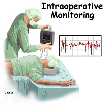
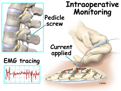
Whenever we are giving strengthening exercises to the patients, we continuously give such encouraging commands to the patient to enable them perform better each day. This in itself is a feedback. When a patient performs a muscle contraction, electric energy is produced which is recorded by our electrodes and displayed on the screen. Hence patient can see and hear his own muscle performance. And since it involves a biological tissue, it is called biofeedback.
Using the same feedback for training a muscle using electromyography is Electromyography Biofeedback or EMG biofeedback. This doesnt mean this is too difficult a concept to understant and implement. All it needs is out of the box thinking and actually applying our learnt theory in the practical scenario.
Recent research evidences give controversial results, rather detrimental results regarding giving electrical stimulation for muscle strengthening. In such case, what is left for us to train such muscles? Are only passive movements and active movements enough for rehabilitation? Can it give patient satisfaction? Rather than giving a controversial treatment just to satisfy the patient, why not think different and think new?
The same equipment that performs NCV and EMG studies can be used for this purpose. I know this leaves many of the readers in surprise. But simple. If we can see Motor Unit Action Potentials and Interferential Pattern of EMG studies in an equipment, why cant we use the same computer screen sequentially to see the progress of the patient, to let the patient see his progress and in a way get a feedback? Logical. Isn't it? If needle EMG study can be interpreted on the clinical EMG machine for diagnostic purpose, why can't we perform surface EMG for therapeutic purpose to train the muscle? This is EMG biofeedback. No one can create any controversy over this use of this equipment.
HOW TO SEE THE SCREEN

The above figures take us back to our childhood days when we used to do maths. This reminds us of the graph paper wherein each square is one centimeter square area with each connecting horizontal and vertical line one centimeter. We used to adjust X-axis and Y-axis and plot a graph and actually enjoy doing it. We had to decide a unit like X-axis, 1cm= something and Y-axis 1cm=something and then plot a graph.

Now look at the above figure. Though it looks the same as the graph chart, is not our childhood graph. But it is the screen of the EMG equipment we use in clinical set up. They are so identical. Isn't it? Adjustments are also identical. The way we adjust X axis and Y axis in the graph, for the EMG screen, we adjust the horizontal screen and the vertical screen. Irrespective of using it for MUAP analysis or Biofeedback training, basic screen adjustments remain the same.The vertical line stands for the amplitude or the strength of contraction as measured in microvolts or millivolts depending upon patient's need. The horizontal line stands for time as measured in milliseconds. We can adjust the amplitude in the graph known as GAIN ( amplitude/cm) and time known as SWEEP ( sweep speed/sec)
In a graph, increasing or decreasing the units of X or Y axis changes the size of the graph. Similarly increasing or decreasing the gain and sensitivity will change the size of the graph seen on the EMG computer screen. This can be used as a feedback to train the patient.
While training a muscle, we adjust a gain. We tell the patient to perform the muscle action and see how much is the total amplitude of that action is. This gives us an idea of patient's capability of performance. We adjust the threshold accordingly. THRESHOLD is the maximum limit which we aim the patient should be able to do at that particular time. And we instruct the patient to perform the activity such that the MUAP crosses the threshold line

Above figure is an example of one such patient scenario graph. Red graph is the MUAP when a patient performs muscle contraction. "2" which is seen on Vertical axis can be considered as Threshold and we instruct the patient to contract still strongly so that the red graph crosses the horizontal line passing through "2". Patient seeing the electric conversion of his own contraction as a graph performs still stronger contraction and ultimately puts in efforts to reach "2".
Progression can either be made by telling the patient to reach "3" or by increasing the gain. Once we increase the gain, graph size will be smaller and then once again we can instruct the patient to reach"2". On seeing the graph smaller, patient will definitely remember previous size and put in efforts to reach the previous day's target because patients know target and not the GAIN values. This is how progression made without any documented contraindications.
The same can be used to relax over active muscle aswell. Just reverse the sequence. Tell the patient to bring down the activity level, relax so that the graph falls below the threshold and progressed by reducing the gain.
ELECTRODES USED -
Surface electrodes are used. Two surface electrodes placed over the muscle to be trained and a Ground electrode used for earthing. This avoids all controversies, conflicts and quarrels. Instrumentation is same which is not at all expensive. All it needs is just innovative thinking and an inclination to do something good.
Rotator cuff partial tear patients often present with scapular dyskinesis with hyperactive upper trapezius with affect pure glenohumeral flexion and abduction. We have achieved very good results in our set up training such patients with EMG biofeedback using our Clinical EMG NCV equipment. We focused on training relaxation of upper trapezius and training rotator cuff. Results are excellent
We have a notion that EMG biofeedback training needs expensive and glamorous instrumentation which is seen only in western countries. Our conventional EMG/NCV equipment can do the same work. All it needs is our inclination, our WILL to do and OFCOURSE KNOWLEDGE OF BASICS OF EMG, INSTRUMENTATION OF EMG AND EMG BIOFEEDBACK
This blog is just the basics of EMG biofeedback. Discussion and description can be interestingly endless. But even this much of basic inculcation can be a small seed sown for the future new era of muscle diagnostics and training


